


ARC-Grain Crops, Potchefstroom

Oriah Lekhetho Maffa,
University of Pretoria, ARC-Grain Crops, Potchefstroom

Yvonne Maila,
ARC-Grain Crops, Potchefstroom
Maize producers are faced with an increasing number of disease threats that significantly reduce yield and grain quality, leading to an impact on trade. The early identification of diseases is therefore an essential step in developing control strategies for any pathogen. Among these current threats are Goss’s wilt, which has officially been reported for the first time in South Africa during 2025.
This disease is caused by Clavibacter michiganensis subsp. nebraskensis (Cmn) and has since become an important disease in several maize-producing regions of the country. However, field symptom identification of Goss’s wilt can easily be confused with that of other foliar diseases, herbicide injury, or environmental stress symptoms. Advances in diagnostic technology allow for rapid and reliable identification of the pathogen. To assist producers and company agents, this article highlights key diagnostic techniques that are available for Goss’s wilt, ranging from simple field observations to advanced molecular techniques.
Diagnostic methods that can be used to identify Goss’s wilt
Field diagnosis; the first line of detection
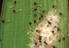
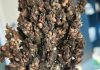
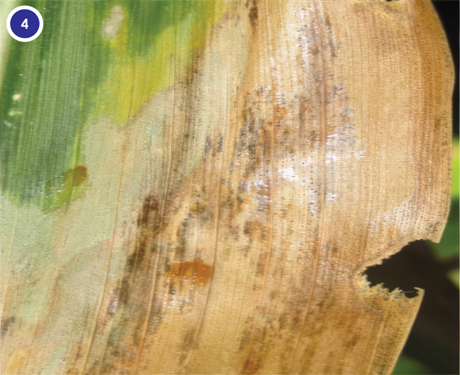
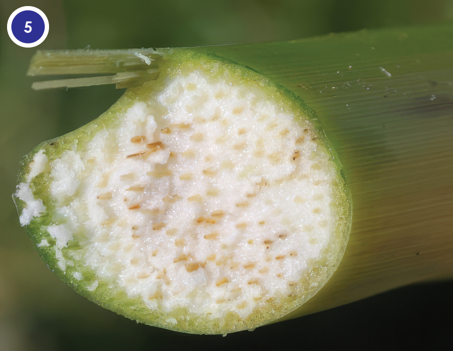
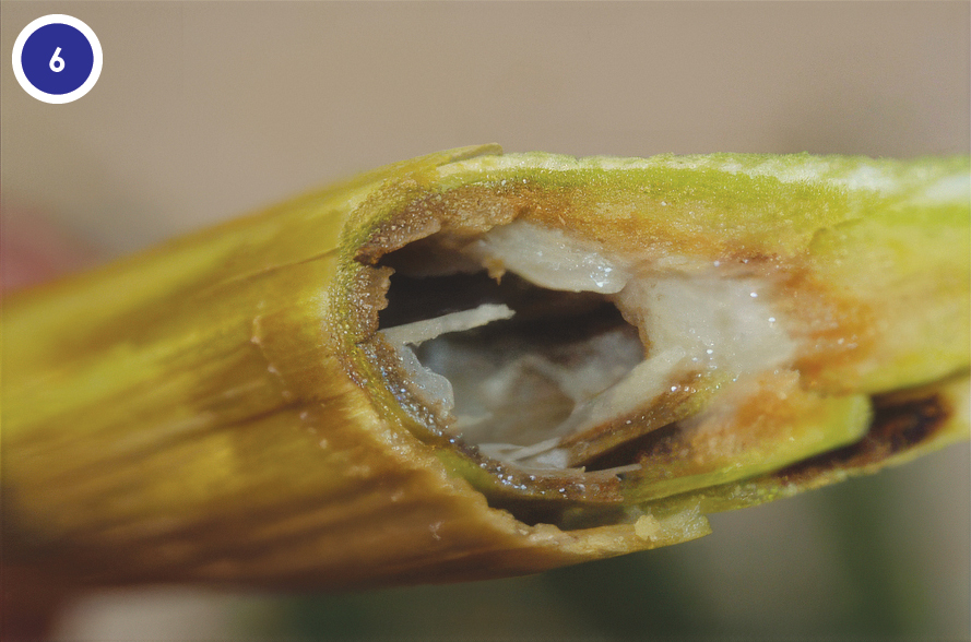
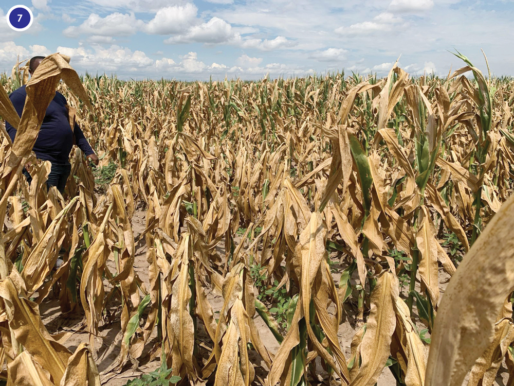
Field diagnosis is an important step where plants are inspected for characteristic symptoms. Goss’s wilt symptoms typically manifest as long grey-green to tan leaf lesions with dark green to black, water-soaked margins and shiny bacterial exudates (also known as freckles). Advanced systemic infections are marked by internal orange-to-brown stalk discolouration, wilting, stunting, collapse of infected plants and, in severe cases, death of the plant (Photo 1-7) (Jackson et al., 2007; Osdaghi et al., 2023).
Although this method is a rapid, inexpensive way that can be performed directly in the field, these symptoms alone cannot confirm Goss’s wilt. First and foremost, these symptoms may also resemble those of other foliar maize diseases or abiotic stress. Field diagnosis will, in addition, not be successful in the case of latent or non-symp-
tomatic infections.
Microscopic observations
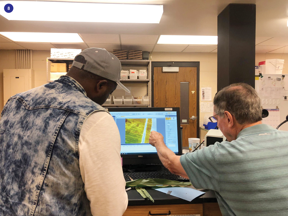
In the laboratory, one of the first Goss’s wilt disease checks is to look for bacterial strea-
ming. A small piece (1 cm2) of leaf is taken from the edge of a lesion and placed on a clean glass microscope slide. A single drop of clean water is added and a coverslip placed over the drop of water. This slide is then examined under a compound micro-
scope, starting from the lowest magnification to the highest (40x to 100x).
However, while this method is useful to identify the presence of a bacterial infection, it cannot confirm the bacterium as Clavibacter nebraskensis. Laboratory tests such as polymerase chain reaction (PCR) are then conducted to confirm the presence of the Goss’s wilt pathogen.
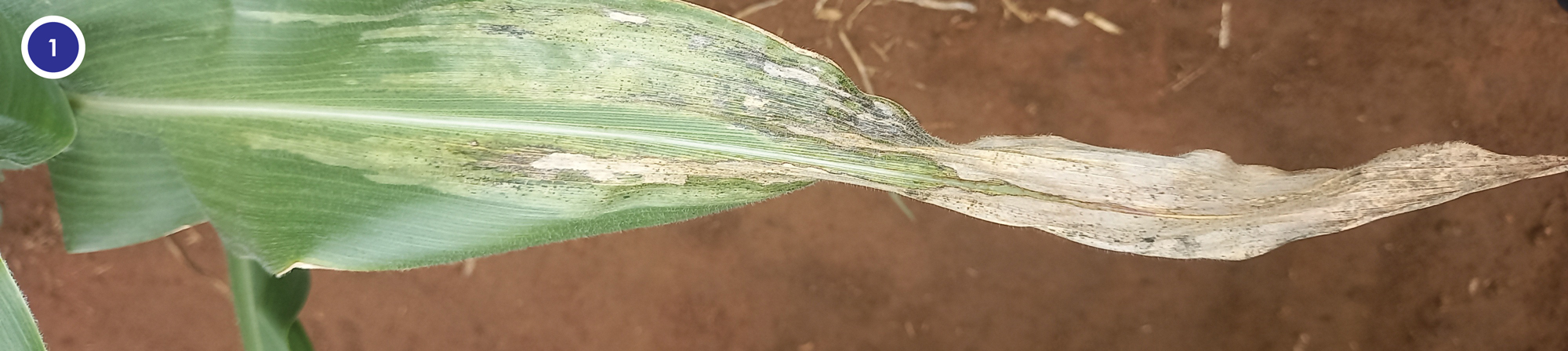
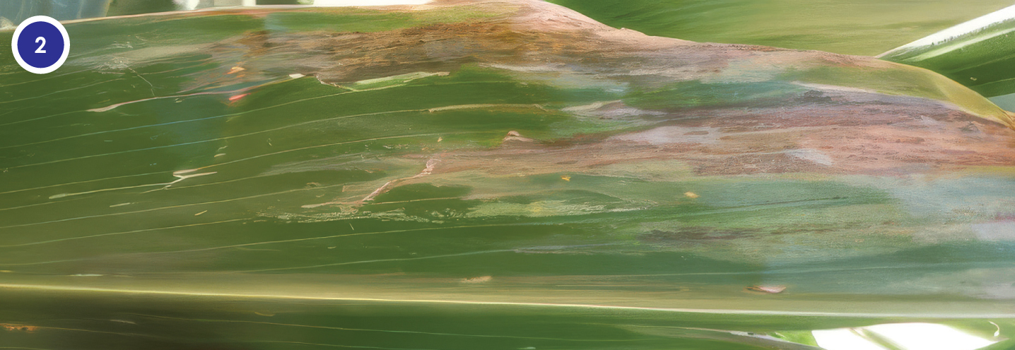
and tissue decay. Photo: Dr Henry Njom, ARC-Grain Crops
Rapid field tests
Rapid tests are available for on-site detection of various Clavibacter subspecies, most notably Clavibacter michiganensis subsp. michiganensis (Cmm) that causes bacterial canker in tomatoes. Common antigens are shared among all C. michiganensis subspe-
cies, which enables us to use this assay for a presumptive detection of C. michiganensis subsp. nebraskensis.
The advantage of this test lies therein that it can provide preliminary identification results within 30 minutes without specialised equipment or training. However, cross-reactions with non-Clavibacter bacteria can occur, which renders this assay less ideal for definitive routine identification of the Goss’s wilt pathogen.
Culture-based diagnosis
Traditional microbiology methods remain important for disease diagnosis, maintaining pure reference strains and verifying pathogenicity through Koch’s postulates to prove that the organism caused the observed symptoms. Like with other coryneform phytopathogenic bacteria, it is possible to isolate C. nebraskensis on enriched media such as yeast extract peptone glucose agar (YPGA) and nutrient broth-yeast extract (NBY). In 1979 Gross and Vidawer developed a selective agar medium known as
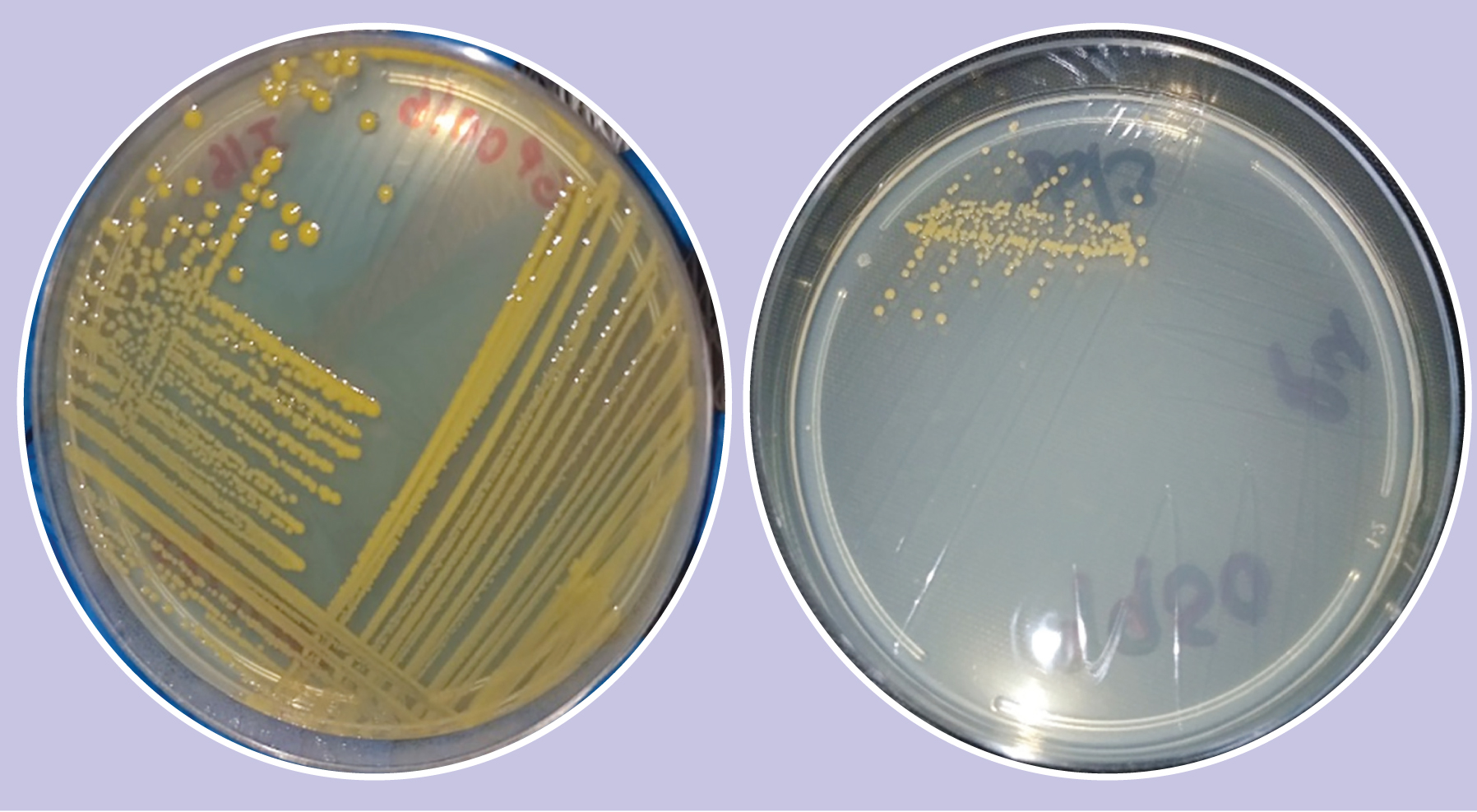
C. nebraskense selective (CNS) medium. CNS is used for the isolation of C. nebraskensis from maize tissue and soil. Small, surface-sterilised pieces (1 cm2) of symptomatic leaf tissues can be plated on these various media and incubated at 28 °C for four to seven days. After six days of growth on YPGA, NBY, or CNS media, typical colonies of C. nebra-
skensis appear yellow or apricot-orange, round, convex, glistening, and 4 mm in diameter.
Molecular-based diagnostics
Molecular-based assays for C. michiganensis subspecies differentiation are available. However, it may be impractical for many plant disease diagnostic laboratories to perform as routine sample processing.
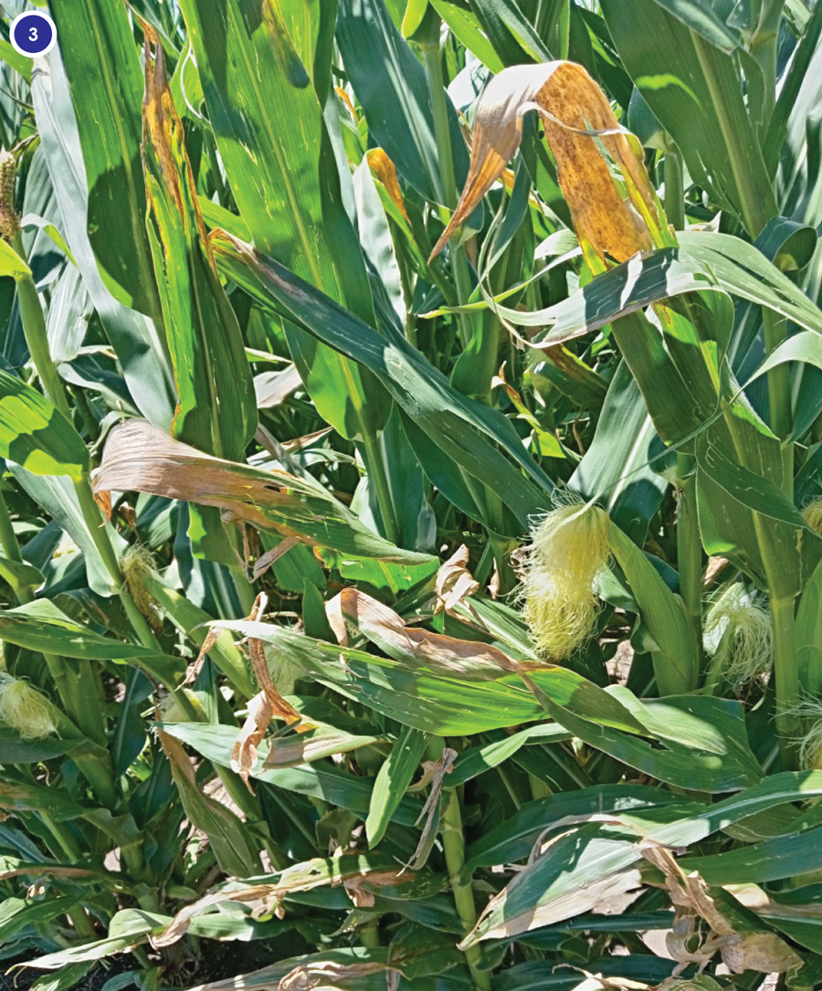
Photo: Yvonne Maila, ARC-Grain Crops
Rapid DNA amplification tests
This technique uses the isothermal nucleic acid amplification technique such as loop-mediated isothermal amplification (LAMP) (Yasuhara-Bell et al., 2016). This rapid molecular diagnostic method enables the detection of pathogen DNA without the need for sophisticated laboratory thermocyclers. Unlike conventional PCR, nucleic acid is amplified at a constant temperature (approximately 39 to 65 °C) using a simple portable device such as the AmplifyRP XRT (Agdia), which is particularly well suited for field diagnostics and point-of-care testing. LAMP can detect pathogen DNA within 30 to 60 minutes, providing a rapid, sensitive, and cost-effective means of diagnosing Goss’s wilt.
The fast turnaround time allows producers, extension officers, and crop advisors to make timely and informed disease management decisions before the infection spreads further. Because the technology is easy to use, requires minimal equipment, and can be operated outside a conventional laboratory, it is especially valuable for on-site disease surveillance and early detection.
Polymerase chain reaction (PCR)
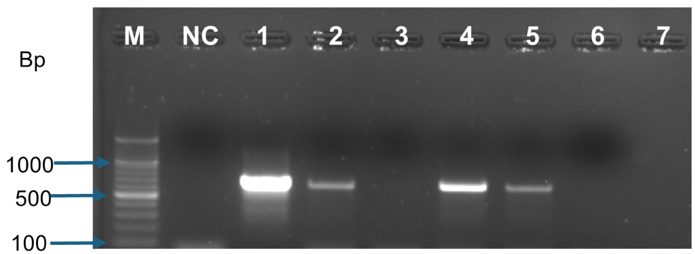
Modern DNA-based laboratory tests such as PCR provide fast and accurate detection of C. nebraskensis. Over the years, researchers have developed sensitive PCR methods that can detect the pathogen in maize seed and infected plants using very small amounts of bacterial DNA. Advanced real-time PCR (qPCR) techniques also measure the population density of the pathogen, enabling researchers and diagnostic laboratories to confirm infections rapidly and reliably.
Primers exist for identification and quantification of C. nebraskensis such as the PSM1 and CM3 (Ayala-Labarrios et al., 2004), CmnFP-OUTER/CmnRP-OUTER and CmnFP-rNNER/CmnRP-INNER (Feng et al., 2004), and CMN_01184 (McNally et al., 2016). These molecular tools play a crucial role in accurate, early diagnosis, disease monitoring, and supporting of effective management decisions.
Diagnostic support and call for collaboration
Plant disease management relies on rapid, accurate, and field-based identification techniques. Combining field scouting with modern detection tools, maize producers can make informed management decisions that protect yield, profitability, and biosecurity. When Goss’s wilt is suspected, visual symptoms should always be confirmed through laboratory testing.
The Agricultural Research Council-Grain Crops in Potchefstroom offers diagnostic testing of plant samples and encourages collaboration among producers, researchers, and extension officers to improve early detection, reporting, and monitoring of Goss’s wilt. These efforts will strengthen biosecurity and help protect maize production in South Africa.
 For further information or to arrange for sample collection, please contact Dr Henry Njom at 018 299 6225 or njomh@arc.agric.za.
References
- Ayala-Labarrios, LA, Rodríguez-Herrera, R & Aguilar-González, CN. 2004. Detección de Clavibacter michiganensis subsp.
nebraskensis (Schuster, Hoff, Mandel y Lazar) Vidaver y Mandel, usando la reacción en cadena de la polimerasa. Revista Mexicana de Fitopatología, 22, 239-245. - Feng, L, Yu, D, Wang, Y, Wu, C, Wu, X, Zhang, J et al. 2014. Nested-PCR detection of Clavibacter michiganensis subsp. nebraskensis. Journal of Food Safety and Quality, 5, 3933-3938.
- Gross, DC & Vidaver, AK. 1979. A selective medium for isolation of Corynebacterium nebraskense from soil and plant parts. Phytopathology, 69, 82-87.
- Jackson, TA, Harveson, RM & Vidaver, AK. 2007. Reemergence of Goss’s wilt and blight of corn to the central High Plains. Plant Health Progress, 8, 44.
- McNally, RR, Ishimaru, CA & Malvick, DK. 2016. PCR-mediated detection and quantification of the Goss’s wilt pathogen Clavibacter michiganensis subsp. nebraskensis via a novel gene target. Phytopathology, 106, 1465-1472.
- Osdaghi, E, Robertson, AE, Jackson-Ziems, TA, Abachi, H, Li, X & Harveson, RM. 2023. Clavibacter nebraskensis causing Goss’s wilt of maize: five decades of detaining the enemy in the New World. Molecular Plant Pathology, 24(7), 675-692.