


Dr Lisa Rothmann,
Department of Plant
Sciences, Plant
Pathology, University
of the Free State

Mariana van Deventer, PhD researcher, Plant Pathology, Stellenbosch University

Kwanele Sabela,
Department of Plant
Sciences, Plant Pathology, University of the Free State

Dr Diane Mostert, chief technical officer, Plant Pathology, Stellenbosch University

André van der Merwe, chairman, Industrial
Engineering, Stellenbosch
University
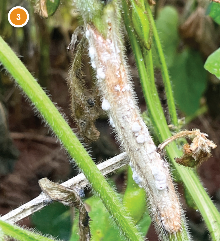
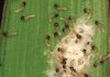
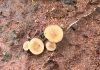
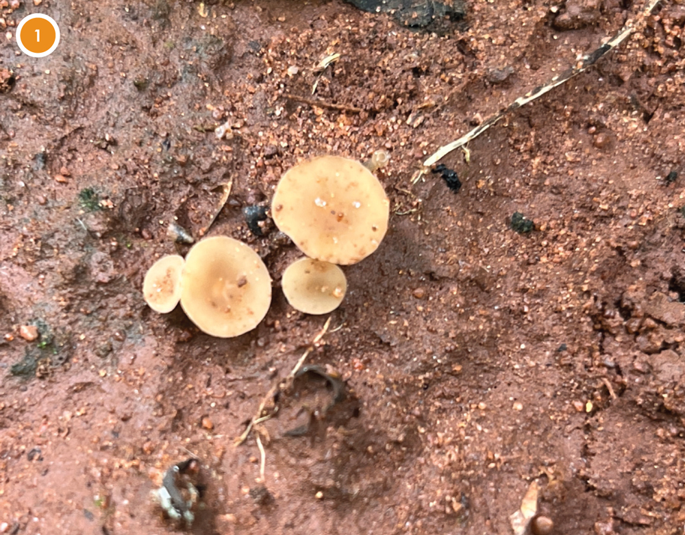
One of the major challenges in managing diseases caused by Sclerotinia sclerotiorum is that much of the infection risk begins before symptoms are visible. The pathogen produces microscopic airborne spores, called ascospores, which are released from apothecia and move through the crop canopy (Photo 1).
Apothecia are the small mushroom-like structures produced by sclerotia when soil and canopy conditions are favourable. Understanding when ascospores are present and where they move is an important step towards improving disease monitoring, predicting disease risk, and making informed management decisions.
Canopy closure helps maintain the cool, humid microclimate needed for apothecial development. In soybean, this often coincides with the flowering stage, although the timing depends on planting date. This is also the crop’s most susceptible period, when mature apothecia may release ascospores that can initiate infection. An apothecium may continue to produce and release spores for more than a week under favourable conditions, with spores released in successive ‘puffs’ rather than single events and decreasing per apothecium as it approaches maturation.
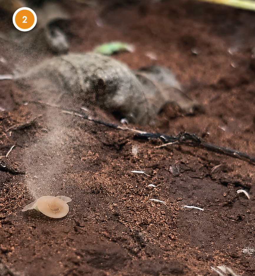
The puff forcibly ejects ascospores a few centimetres from the apothecium, after which they are carried by air and turbulence through and above the crop canopy, sometimes for a few kilometres. The apothecia release spores both during the day and at night, depending on changes in temperature, relative humidity, and air movement. There is no single, universally applicable release window, although in one study, the peak time for available ascospores was observed between 8 and 13 hours of the day. Instead, these releases are governed by favourable environmental conditions, thereby influencing apothecial behaviour and ascospore release.
Ascospores remain viable in the air for about 17 hours, provided that the relative humidity is above 60%. This survival period is reduced under unfavourable conditions such as high temperature, drying, and exposure to harsh UV radiation. Under cool, protected laboratory conditions, around 5 to 10 °C and moderate relative humidity, ascospores may remain viable for two weeks or longer.
However, once deposited on exposed plant surfaces in the field, viability declines much faster, where only half of the ascospores remain viable after two days, less than a quarter after four days, and less than 10% after six days. After a two-week period, very few viable ascospores are expected to remain, especially where spores are exposed to temperatures of 25 °C or higher, drying conditions, and direct sunlight.
Spore sampler
As part of the #SclerotiniaZA research network, the authors recently had the opportunity to test a newly developed wind-powered spore sampler in a soybean field site. This spore sampler was designed and is currently being used within canola stem rot research at Stellenbosch University under the supervision of Dr Diane Mostert.
The work forms part of the PhD research of Mariana van Deventer, who is developing and applying the sampler in collaboration with Prof André van der Merwe from Industrial Engineering. Dr Lisa Rothmann from the University of the Free State is involved as co-supervisor on van Deventer’s PhD project. The soybean field test described here, therefore, represents a collaborative opportunity to explore how this tool may also help researchers better understand the presence of ascospores in other crop systems.
The sampler was tested under field conditions where Sclerotinia apothecia were actively present. The rods of the sampler were exposed from 10:00 to 16:00 in a field heavily infested with apothecia. After this, the sampler was moved to a second field where no apothecia were observed and left there from 16:00 until 09:00 the following morning. The rods of the sampler act as a surface on which airborne spores can be captured as they move through the crop canopy. After exposure in the field, DNA is extracted from the rods and analysed using a specialised molecular technique called quantitative PCR, or qPCR. This technique allows researchers to detect Sclerotinia DNA and quantify how many copies of the target DNA are present.
Molecular diagnostics using qPCR are highly accurate and specific for detecting DNA; imagine finding a needle in a haystack with a powerful magnet, where the haystack represents a DNA sample, the needle represents a unique part of the pathogen’s genetic code, and the magnet represents the qPCR reaction. Once the pathogen DNA is detected in the reaction, the qPCR machine records a fluorescent signal, indicating a positive sample. There may be more than one needle (the target copy of DNA) present in the haystack (the sample); the more copies are detected, the stronger the signal is, and the quantity of target DNA copies can be calculated. Based on qPCR results, it is possible to determine whether and when the pathogen was present or absent in the environment, and to compare DNA levels to identify when more or less DNA was present.
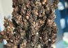
The ability of ascospores to cause infection can be supported by field observations, such as the presence of apothecia (Photo 2), which may indicate active local ascospore production and therefore a higher risk of nearby exposure, or the development of Sclerotinia disease in the sampled field at a later stage. This can be investigated more directly through petal tests, in which petals exposed to ascospores are assessed for pathogen infection. Importantly, ascospores were detected during both sampling periods, indicating that apothecia can actively release ascospores during both the day and night and that these spores may be transported through the local atmosphere. See the accompanying video for the actual event captured in our soybean field here.
Practical research
Although this was a small field test, it is an exciting example of how practical research tools can help researchers better understand disease risk under real production conditions. It should therefore be viewed as a first field demonstration rather than a complete risk system. Spore sampling can provide valuable insight into when spores are present in the field, how they move through the crop canopy, and how this information may eventually support more informed disease management decisions for producers.
This work also reflects the value of collaboration within the #SclerotiniaZA network, where researchers working on different crops and disease systems can share tools, field sites, and expertise to build a stronger understanding of Sclerotinia diseases in South Africa. By combining field observations, engineering design, and molecular detection, more precise questions can be asked about when ascospores are present and how this information can eventually support disease management. In the future, the authors look forward to sharing more about the origin of these spore traps and the important canola stem rot research being conducted at Stellenbosch University.